low end tidal co2 pulmonary embolism
The diagnosis of pulmonary embolism is often missed. The authors aimed to define the optimum ETCO2 to conclusively exclude a pulmonary embolic event.

A Low End Tidal Co2 Arterial Co2 Ratio During Cardiopulmonary Resuscitation Suggests Pulmonary Embolism Resuscitation
This study however aimed to predict or exclude PE using the end-tidal carbon dioxide ETCO 2 value and alveolar dead space fraction AVDSf together.

. 1 that etCO2 can dist. The diagnosis of pulmonary embolism PE because of nonspecific clinical presentation remains as a challenge for emergency physicians. In thromboembolism ETCO2 is significantly lower than normal due to the reduction of pulmonary perfusion and increased alveolar dead space that reduces the amount of CO2 exhaled from the lungs so venous carbon dioxide pressure PvCO2 increases and all of these changes lead to an increase in arterial CO2-ETCO2 gradient.
ETCO2 end-tidal carbon dioxide. 2 There have been reports of carbon dioxide emboli occurring in various procedures including laparoscopic. Pulmonary embolism increases alveolar dead space resulting in low end-tidal CO 2 EtCO 2 relative to arterial CO 2 PaCO 2 tension.
In a similar larger study of 298 patients Hemnes et al. Symptoms demographic date Wells score D-dimer levels and the gold standard computed. The aim of this study was to investigate the value of end-tidal carbon dioxide ETCO2 and alveolar dead space fraction ADSF in the diagnosis of PE.
The diagnosis of pulmonary embolism is often missed. Pulmonary thromboembolism results in dead space ventilation and therefore prevents meaningful gas exchange in the subtended lung unit yielding an alveolar CO 2 content as low as 0. This was a prospective diagnostic study set in a British acute medical admissions ward.
Decreased perfusion to the lungs and reflex hyperventilation to compensate decreases end-tidal CO2 or ETCO2 5. One hundred consecutive patients with suspected pulmonary embolisms PEs were enrolled over 6 months in 2012. This may result from such ventilatory problems as high mean airway pressure or inadequate exhalation time resulting in overdistention or from such circulatory problems as.
The diagnosis of pulmonary embolism is often missed. Pulmonary embolism increases alveolar dead space resulting in low end-tidal CO2 EtCO 2 relative to arterial CO 2. End-tidal CO2 ETCO2 can represent dead space ventilation.
Several studies have reported that computed tomography pulmonary angiography is the best method for diagnosing pulmonary embolism PE. Pulmonary embolism PE is a frequent health problem representing a diagnostic challenge with high mortality and morbidity rates. Massive pulmonary embolism PE results in low CO 2 transport due to hemodynamic compromise together with an alveolar dead space effect increase in poorly perfused but well ventilated lung areas.
Because pulmonary embolism obstructs pulmonary blood vessels not the airways. Thus a low EtCO 2 PaCO 2 ratio during resuscitation may be a sign of pulmonary embolism. CTPA computed tomography pulmonary angiogram.
Dead-space ventilation results in ventilated alveoli with insufficient perfusion which leads to low ETco 2. A Low End Tidal Co2 Arterial Co2 Ratio During Cardiopulmonary Resuscitation Suggests Pulmonary Embolism Resuscitation Capnography Provides Bigger Physiological Picture To Maximize Patient Care Jems Ems Emergency Medical Services Training Paramedic Emt News Different Capnography Traces A Sudden Drop In E 0 Co2 B Download Scientific Diagram. At a cut-off of 36 mmHg capnography achieved a negative predictive value of 966.
Riaz I Jacob B Pulmonary embolism in Bradford UK. End-tidal carbon dioxide tension P ETCO 2 is a physiological surrogate for vascular obstruction from PE. A DVT Is A Blood Clot Which Can Travel To The Lungs And Lead To A PE.
Carbon dioxide embolism is a rare but potentially serious complication of laparoscopic procedures. Thus a low EtCO 2 PaCO 2 ratio during resuscitation may be a sign of pulmonary embolism. ETCO2 levels of patients with suspected PE.
Continuous pulmonary arterial pressure can be used to evaluate for gas embolism. All study participants underwent end-tidal CO 2 determination within 24 h of state-of-the-art diagnostic imaging. Pulmonary embolism PE is associated with approximately 100000 deaths per year in the United States and the incidence of deep vein thrombosispulmonary embolism in the United States is estimated at more than 350000 cases annually.
1 It is caused by entrapment of carbon dioxide in an injured vein artery or solid organ and results in blockage of the right ventricle RV or pulmonary artery. Ad Early Medical Attention May Help Reduce The Chance Of DVT Or PE Becoming More Serious. 2014 Apr14 2 Objective.
Post hoc analysis of data from two porcine studies comparing. End tidal CO2 is reduced during hypotension and cardiac arrest. Role of end-tidal CO2 as a screening tool Clin Med.
This figure shows that no patient with an ETCO2 3253 had a pulmonary embolism. Sudden decrease or loss of end-tidal CO2 suggests a drastic decrease in cardiac output due to gas embolism. Underwent ETCO2 determination within 24 hours of diagnostic imaging 19.
To evaluate whether EtCO2 measurement can be used at the bedside to exclude pulmonary embolism PE. We investigated the effect of massive pulmonary embolism MPE on end tidal CO2 etCO2 and tested two hypotheses. Pulmonary embolism increases alveolar dead space resulting in low end-tidal CO 2 EtCO 2 relative to arterial CO 2 PaCO 2 tension.
End-tidal clearance must be evaluated in the context of the patients perfusion status. Ad Get Info On An Rx Option To Treat Lower The Risk Of Recurrent DVTPE Blood Clots. Cardiac arrest from PE is associated with extremely low etCO2 readings during CPR.
Several studies have reported that computed tomography pulmonary angiography is the best method for diagnosing pulmonary embolism PE. Arterial to end-tidal partial pressure of carbon dioxide Pa-Et CO 2 gradient may be useful in the evaluation of PE. 1 The diagnosis of pulmonary embolism poses a diagnostic challenge in the emergency department ED despite.
In the group of patients finally diagnosed with PE n 39 end-tidal CO 2 was significantly lower than in the group without PE or in healthy volunteers. The line represents a threshold of 3253 mmHg. Visit The Official Patient Site For Additional Information About DVTPE Blood Clots.

Figure 1 From Pulmonary Embolism Pathophysiology Diagnosis Treatment Semantic Scholar
Detection Of Pulmonary Air Embolism Capnography

Capnography Capnography

Capnography Provides Bigger Physiological Picture To Maximize Patient Care Jems Ems Emergency Medical Services Training Paramedic Emt News

Basics Of Waveform Capnography Waveform Capnography Grepmed

Understanding The Arterial To End Tidal Co2 Gradient P A Et Co2

Waveform Capnography In The Intubated Patient Emcrit Project

Capnography And Pulmonary Embolism Chapter 21 Capnography

Basic Capnography Interpretation Nuem Blog

Pre Hospital Capnography Ppt Download
Physiology Capnography
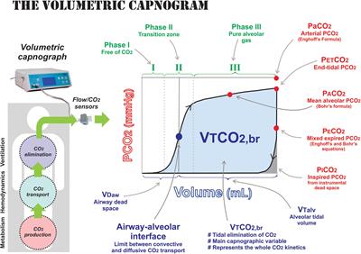
Frontiers Monitoring Expired Co2 Kinetics To Individualize Lung Protective Ventilation In Patients With The Acute Respiratory Distress Syndrome Physiology

Different Capnography Traces A Sudden Drop In E 0 Co2 B Download Scientific Diagram

Pulmonary Embolism Alters Alveolar Dead Space And End Tidal Carbon Download Scientific Diagram

Evaluation Of Suspected Pulmonary Embolism Utilizing End Tidal Co2 And D Dimer The American Journal Of Surgery

Flow Diagram Of The Study Pet Co2 End Tidal Carbon Dioxide Tension Download Scientific Diagram

A Low End Tidal Co2 Arterial Co2 Ratio During Cardiopulmonary Resuscitation Suggests Pulmonary Embolism Resuscitation

Bedside End Tidal Co2 Tension As A Screening Tool To Exclude Pulmonary Embolism European Respiratory Society

Interpreting Capnograms Rk Md